Foraminal Stenosis
Foraminal stenosis — also called neural foraminal stenosis or foraminal narrowing — occurs when there is a narrowing in parts of your spine, which may compress your spinal nerves and cause nerve issues and pain. Rojeh Melikian, M.D., has experience treating patients with this condition using minimally invasive surgical techniques.
What Is Foraminal Stenosis?
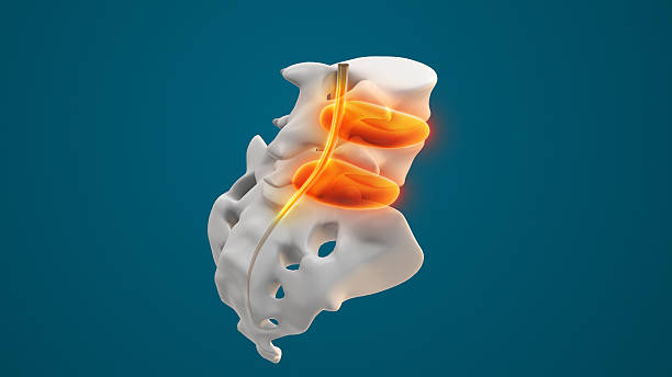
Foraminal stenosis is a type of spinal stenosis characterized by a narrowing of the neural foramina — the openings on both sides of the spinal vertebrae. Spinal nerves exit the spine through neural foramina. If these openings are too narrow, they put pressure on the nerves, causing nerve pinching or pain.
Foraminal stenosis can impact several regions of the spinal column. Lumbar foraminal stenosis affects the lower back and is the most common type. Another common type is cervical foraminal stenosis, which occurs in the cervical spine.
Foraminal stenosis is different from central stenosis. Neural foraminal stenosis is the narrowing of the intervertebral foramen where spinal nerves exit the spine, while central stenosis is the narrowing of the spinal canal.

Foraminal Stenosis Symptoms
Some patients with foraminal stenosis do not have symptoms. However, those with severe narrowing can experience symptoms like:
- Numbness: Nerve root compression can cause localized numbness.
- Paresthesia: When your nerves are pinched, you may feel a tingling, pins-and-needles sensation in the areas the affected nerve supplies.
- Pain: Electric, sharp or burning pain may radiate throughout your limbs.
- Muscle weakness: Affected limbs can have weakened muscles.
Causes of Foraminal Stenosis
Various preexisting conditions and risk factors can cause neural foraminal stenosis, including:
- Degenerative disc disease: Moisture loss can cause spinal discs to degenerate over time. As the discs flatten, the foraminal opening shrinks.
- Osteophytes: Bone spurs and other overgrowths can project into the foraminal space, narrowing it.
- Facet joint arthritis: Arthritis in the joints at the back of the spinal segments can lead to thickening, reducing the foraminal space.
- Herniated or bulging discs: When disc material herniates or ruptures, it can compress nerve roots in the foramen.


How Are Osteophytes on the Spine Diagnosed?
If you’re experiencing bone spur symptoms, your orthopedic spine surgeon will perform a physical examination, then follow up with imaging tests. An X-ray of your spine confirms the presence and location of osteophytes. An MRI can evaluate spinal discs and nerves, while a CT scan shows bone growths in greater detail.
How Is Foraminal Stenosis Diagnosed?
Foraminal stenosis can be diagnosed based on an evaluation of your symptoms. Each type of foraminal stenosis causes symptoms in different areas, so a specialist can use the affected dermatome to locate the involved spinal segment.
If you aren’t experiencing symptoms, or your doctor wants to confirm the diagnosis, a magnetic resonance imaging (MRI) test will provide detailed images. A computed tomography (CT) scan with a myelogram is also an option if you cannot undergo an MRI.


How to Prevent Bone Spurs in the Neck and Back
Since bone spurs can develop as you age, they cannot be wholly prevented. However, you can take some steps to delay or reduce the wear and tear on the spine that causes bone spurs:
- Get adequate nutrition to keep your tissues strong and prevent osteoporosis.
- Do low-impact exercises like cycling or swimming to strengthen your musculoskeletal system and take stress off your spine.
- Maintain a good posture to avoid putting extra stress on your spine.
Schedule a Consultation Today
Rojeh Melikian, M.D., is a board-certified orthopedic spine surgeon who uses microsurgery techniques to treat patients with foraminal stenosis and other spinal conditions. Experience our patient-first care by contacting us online or calling 949-806-6946 to schedule a video or phone consultation. Dr. Melikian can also provide a second opinion on your foraminal stenosis treatment.