In this area you will find a list of some common diagnosis and what they mean. Click on the links below to review each diagnosis in detail. If you have any questions about your particular diagnosis, contact us to schedule an appointment to review it in detail.
Disc Herniation
Introduction
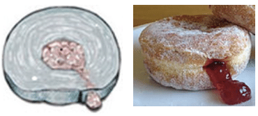
The human spine consists of bones called vertebrae stacked on top of one another in a configuration that supports the weight of your body and protects your spinal cord and nerves. In between each set of bones is a disc which acts as a shock absorber. Each disc is composed of an outer shell of strong material and an inner core of a jelly-like substance called the nucleus pulposus. As a disc ages or becomes damaged due to trauma, you may develop a hole in the outer ring, allowing some of that jelly-like nucleus pulposus to escape and squeeze out. For a quick visualization, imagine what happens when you squeeze a jelly donut and see the jelly squirt out the hole in the side.

Symptoms
Symptoms will vary depending on the location of the ruptured disc. A ruptured disc in your neck may cause pain around your neck, shoulder blade, or running down your arm.. Disc rupture in your low back can cause low back pain and sciatica (buttock, and leg pain that shoots down either leg). Some patients describe it as electricity or feeling like their leg is on fire. Other symptoms include pins and needles or numbness and tingling. More severe disc ruptures can result in weakness in the muscles in your arms or legs as a result of pressure on the nerves controlling those muscles. In extreme cases, they can cause loss of control over your bowel and bladder. Your pain will tend to increase with coughing or sneezing and with sitting down.
How Does it Affect My Nerves?
The herniated disc can affect nerves roots in one of two ways. The first and most obvious mechanism is through sheer pressure. As the disc ruptures and the nucleus pulposus squirts into your spinal canal, it can pin your nerve roots up against the borders of your spinal canal, which is made of bones and tough ligaments, causing physical trauma to the nerve roots. In addition, the chemical make-up of the nucleus pulposus is very irritating to nerves and causes a large inflammatory response when it contacts a nerve root. This explains why even small disc herniations, which do not exert a lot of physical pressure on nerve roots, can still be extremely painful.
How is a herniated disc diagnosed and treated?
In order to make the diagnosis, your doctor will perform a history and physical exam to see if your symptoms are consistent with a ruptured disc. If appropriate, an MRI of your neck or back will show the exact location and size of the herniation.
The treatment goals are to relieve the discomfort and prevent any permanent injury to your nerves. Treatments range from medications and physical therapy to epidural steroid injections or surgery. Depending on the location and size of the disc, surgical intervention may consist of removing only the piece of the disc that has herniated or removing the entire disc.
For a video explanation by Dr. Melikian, please click here.
Degenerative Disc Disease
Introduction
Degenerative disc disease is part of the natural aging process. Similar to how the cartilage in our hips and knees can wear away over time, the same is true for the discs in your neck and back. As the discs wear away, they function less effectively as shock absorbers in your spine. Imaging a tire which has been worn away and is starting to go flat. This is what happens to the jelly core of each disc, also called the nucleus pulposus. As time goes on, it tends to lose its water content and start to dry out. As the disc dries out, it functions less effectively and can cause neck or back pain. It’s important to note that not everyone who has degenerative disc disease will develop symptoms from it.
What Symptoms Can I Expect?
As the disc in your neck or back wear away, the loss of shock absorption can cause neck or back pain as the bones adjacent to each disc feel more force. In addition to pain, stiffness may become more and more of an issue, especially early in the morning. Additionally, as the height of the disc decreases and as the disc bulges out, it may cause narrowing around your nerve roots which can also lead to arm and leg pain.
How Is It Diagnosed?
For patients with neck or back pain from presumed degenerative disc disease, the diagnosis always begins with a visit to go over your medical history and to do a physical examination. This will check your flexibility, area of discomfort as well as your neurological function to make sure all nerves are working as they should be. X-rays will also be ordered at the time of your visit. The x-rays are usually done with you standing up so we can see if there is any narrowing in the space in between the vertebral bodies. While the X-rays only show us the bones of your spine, we can infer how much the disc has worn away by looking at the space in between each set of vertebral bodies.
If the two vertebral bodies are very close together, this is an indicator of degenerative disc disease. Another sign of degenerative disc disease which can be seen on X-ray are bone spurs which occur as the disc collapses and the two bones start to feel more pressure (as there’s no more cushion between the two). In an attempt to decrease the pressure, bones will grow spurs which increase the surface area amongst which the force is distributed. This is similar to how you form bone spurs, also known as osteophytes, in other parts of your body such as your hips and knees.An MRI or CT scan can also be ordered to evaluate your spine more thoroughly.
An MRI will actually show you what the discs look like instead of having to infer that information from an XR or CT scan. Degenerated discs on MRI will have less water signal than the discs around them and will also tend to look flatter. As seen in Figure 1, the disc labeled in red is much flatter and darker than the one labeled in yellow. MRI’s can also show any areas of nerve compression which may be causing associated numbness, tingling, or weakness. The CT scan shows the bony anatomy of the spine in detail and can also be used to look at the space available for the spinal cord and nerve roots.
What Are My Treatment Options?
After a diagnosis of degenerative disc disease has been made, we will go over your treatment options specific to your particular situation. In the absence of arm or leg weakness, treatment options will always begin with conservative treatments such as physical therapy, anti-inflammatories, and rest. Other options to discuss as well are when to add acupuncture or chiropractic treatments into your treatment regimen. If conservative treatment fail to provide relief or you have associated nerve compression causing arm or leg weakness, surgical treatments can be discussed. These can range from a minimally-invasive decompression surgery to neck or back fusion depending on the particular scenario.
Spondylolisthesis
Introduction
Spondylolisthesis is a condition in which one vertebra slips forward in relation to the adjacent vertebrae. The condition can be one you’ve had from birth or developed over time from trauma or degenerative changes. Over time, it can produce narrowing of the vertebral canal and subsequent nerve root symptoms.
What Symptoms Can I Expect and How is it Diagnosed?
The most common symptoms of spondylolisthesis tend to back, buttock and leg pain. The buttock and leg pain is caused by narrowing of the area where the nerves leave the spinal canal. Symptoms tend to be worse when patients stand up and somewhat better with laying down. This is usually because the forward slippage tends to worsen as you stand up and slips back towards normal when you lay down.
Spondylolisthesis can be diagnosed with an X-ray which will show the area of slippage. As can be see in figure 2, the back of the vertebral bodies should form a smooth line. However, down between the two vertebral bodies highlighted in yellow, you can see where there is a step-off in the red line. This is the area of slippage. An MRI will be able to show the amount of nerve compression associated with the spondylolisthesis. However, keep in mind the MRI in this situation will tend to under call the amount of nerve compression as the slippage tends to decrease when you are laying down (most MRIs are done with you laying on your back).
What Are My Treatment Options?
For those without neurological impairment, meaning weakness in their legs as a result of nerve compression, the first line of treatment options consists of conservative measures such as anti-inflammatory medications and physical therapy. Epidural injections are also a treatment option if the buttock and leg pain are particularly bothersome.
Should more conservative measures fail or leg weakness develop, surgical treatments can be discussed. Surgical treatments for spondylolisthesis, and most spine surgery in general, come in two flavors: decompression alone vs. decompression and fusion. The decompression surgery alone involves removing the portions of bone and soft tissue which are pushing on the nerve roots but does not compromise any of the motion. Adding the fusion allows for the restoration of spinal stability by fusing the two slipped vertebrae together. It also allows for a wider decompression of the nerve roots.
Contact us for an appointment to discuss both options and see which is appropriate for you.
Spinal Stenosis
Introduction
Spinal stenosis is a condition in which there is narrowing of the spinal canal which leads to compression of the spinal cord and/or nerve roots. This can occur in any of the 3 regions of the spine (cervical, thoracic, and lumbar) but is most common in the neck (cervical) and low back (lumbar) regions.
The most common cause of spinal stenosis is arthritis. As put more and more miles on your spine, your discs tend to dry out and shrink and size (see section on degenerative disc disease). When this occurs, the space through which your nerves travels becomes smaller. Over time, you may also form bone spurs which encroach into your spinal canal and compress your nerves or spinal cord.
What Symptoms Can I Expect and How is it Diagnosed?
The most common symptoms of spinal stenosis tend to be neck or back pain accompanied by arm or leg pain (depending on which portion of your spine is affected). If your spinal stenosis is in your lumbar spine, patients tend to complain of back, buttock and leg pain which worsens with standing and sitting. It tends to be relieved with sitting, laying down or leaning over. A common scenario is someone who experiences relief of the pain when leaning on a shopping cart. This is because leaning forward tends to open up the passageways for your nerves, which results in a reduction in symptoms.
If your spinal stenosis is in your cervical spine, the symptoms you experience can be different. Most patients will have some neck pain. You may also experience arm pain to accompany it. The difference with cervical stenosis, unlike lumbar stenosis, is that it tends to affect your spinal cord rather than just the individual nerve roots. This can result in balance issues (feeling unsteady on your feet), difficulty coordinating your hands (buttoning shirts, using zippers), or dropping objects out of your hands. In severe cases, both cervical and lumbar stenosis can result in loss of control of bowel and bladder.
Spinal stenosis is typically diagnosed using an MRI. This will show narrowing of the spinal canal where the nerves travel. If you are unable to get an MRI, a CT scan can also show the narrowing of your spinal canal. Figure 3 shows two MRI cross-sections demonstrating a normal spinal canal and one with cervical stenosis. The yellow lines outline the spinal cord itself while the blue lines highlight the edges of the spinal canal (the space available for the spinal cord to float in). On the left panel is a normal canal with plenty of space around the spinal cord. The white area around the spinal cord is filled with cerebrospinal fluid which acts like a watery cushion in which the spinal cord floats. On the right panel is the MRI of a patient with spinal stenosis. As you can see, the spinal cord shape is deformed from an ellipse into a boomerang shape and there is absolutely no cerebrospinal fluid around it. The spinal canal borders (marked in blue) are so narrow, they are causing spinal cord compression.
What Are My Treatment Options?
Depending on the location and severity of your spinal stenosis, several different treatment options are available. For both cervical and lumbar stenosis, treatments can begin with modifying your activities, participating in physical therapy, and/or epidural steroid injections. Should your stenosis be severe enough to cause arm or leg weakness or should conservative treatments fail, you may be a candidate for a minimally-invasive, microscopic decompression or a minimally-invasive decompression and fusion. The treatment choice depends on the location of your stenosis, the severity of the stenosis and whether or not you have other structural issues in your spine which may necessitate a fusion.
